Doctoral researcher students in the Health Sciences at King’s were invited to submit entries for the Public Engagement Competitions (The HSDTC Science Communication Competition and HSDTC Science Image Competition) for a chance to win £300 and be featured on our website and blog.
Five submissions for each competition were shortlisted and published in the brochure for the HSDTC Annual Research Symposium 2021.
This year, the Symposium took place entirely online via Microsoft Teams on the mornings of 29 and 30 April and the prize-winners of the Public Engagement Competitions were announced at an awards ceremony at the close of the event.
HSDTC Science Image Competition 2021
King’s doctoral research students were invited to submit a visual representation of their research. It needed to either convey the entirety of their research or an aspect of it.
We were looking for images which might explore the social impact that their research has on a global, population, or individual scale. They may be a true representation of the subject, or manipulated post-production, such as with false colouring; or they could show their research in a more abstract way.
Images could have shown, for example, the subject matter of their research, such as an image of cells or the brain, or research in action, for example the methods, equipment or facilities used in their research.
The judges were looking for images which are informative, captivating, and which clearly communicate the research being done. Entries could utilise many techniques, such as: photography; microscopy; medical imaging; new and emerging imaging techniques; data visualisation; artistic media.
Even with the added challenge of a limiting year of lockdowns, the students submitted a variety of creative images. The shortlisted submissions are available below, including our winner Lea Lortal, and runner-up, Andrew Doel:
Lea Lortal (winner)
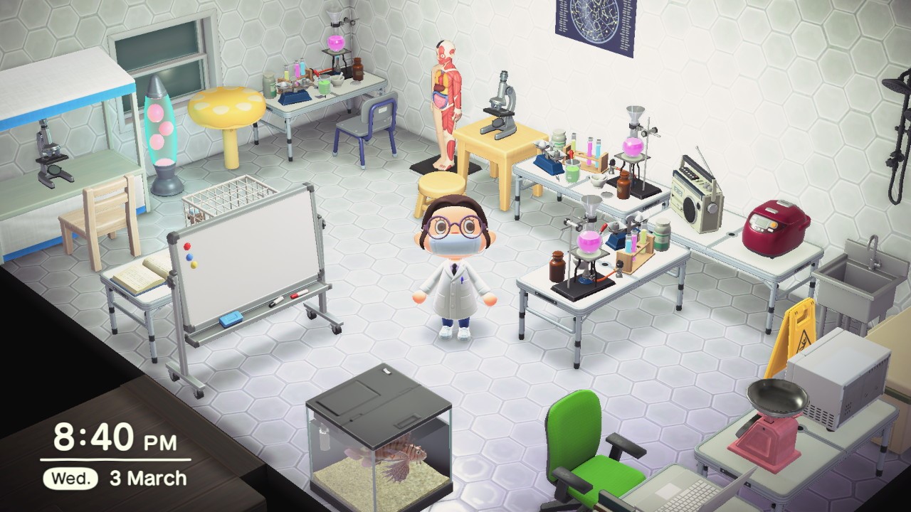
Centre for Host-Microbiome Interactions, Faculty of Dentistry, Oral & Craniofacial SciencesDoing a Lab-based PhD during a pandemic
In 2020-21, research in the lab has been complicated. From March 13th to July 5th, during the first UK lockdown, researchers worked from home, writing reviews and grants, reading as many papers as possible, and planning future experiments. But we still missed the lab. In my free time, I also played Animal Crossing New Horizons, a popular video game where you live on an exotic island and can build your home. On this game, I built my very own research lab and it made me joyful. Now, we’re back in the real lab, which is great, but conditions are more challenging than in the pre-COVID-19 world. Rotations, strict schedules, wearing masks all day and social distancing are part of it. But researchers are still doing their best to advance science and this image, I believe, captures an aspect of current lab research during the COVID-19 pandemic.
Andrew Doel (runner-up),
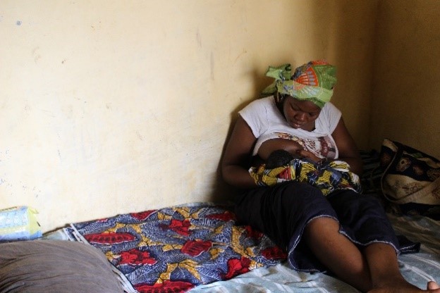
Department of Women & Children’s Health, Faculty of Life Sciences & MedicineNew mother Breastfeeding, The Gambia
The WHO recommends exclusive breastfeeding for the first 6 months postpartum and continued breastfeeding to two years or beyond, owing to its well established benefits for both mother and infant.
Exclusive breastfeeding reduces risk of infant mortality resulting from diarrhoea or other common illnesses. Further, it promotes healthy cognitive development and has been associated with reduced risk of later childhood and adult obesity. It is also beneficial for the mother in helping with child spacing and lessening the likelihood of breast and ovarian cancer.
This picture is of a mother enrolled in The Mothers Infant and Lactation Quality (‘MILQ’) study being conducted in The Gambia. This is her first child, and she has just received lactation counselling – provided as part of the MILQ study whose aim is to create reference values for nutrients and other constituents in human milk. These references will improve current estimates of micronutrient requirements across the first 9 months of lactation. They might also be used as an international reference against which to compare concentrations in different population groups and to evaluate the effects of nutritional interventions, such as maternal supplementation or national fortification programmes on breastmilk quality.
Levels of breastfeeding in The Gambia are good, largely due to it being a cost free form of nutrition for the infant, in a low income context. In recent years, however, urban regions of the country (such as where this picture was taken) have exhibited the beginnings of a ‘Nutrition Transition’, characterised by a move away from traditional diets in preference of westernised foods and altered dietary behaviours. One aspect of this transition is an increase in the use of formula milk and a shift away from WHO recommended practice. This shift highlights the importance and continued relevance of efforts to support adherence to the WHO guidelines.
Cathleen Hagemann (shortlisted)
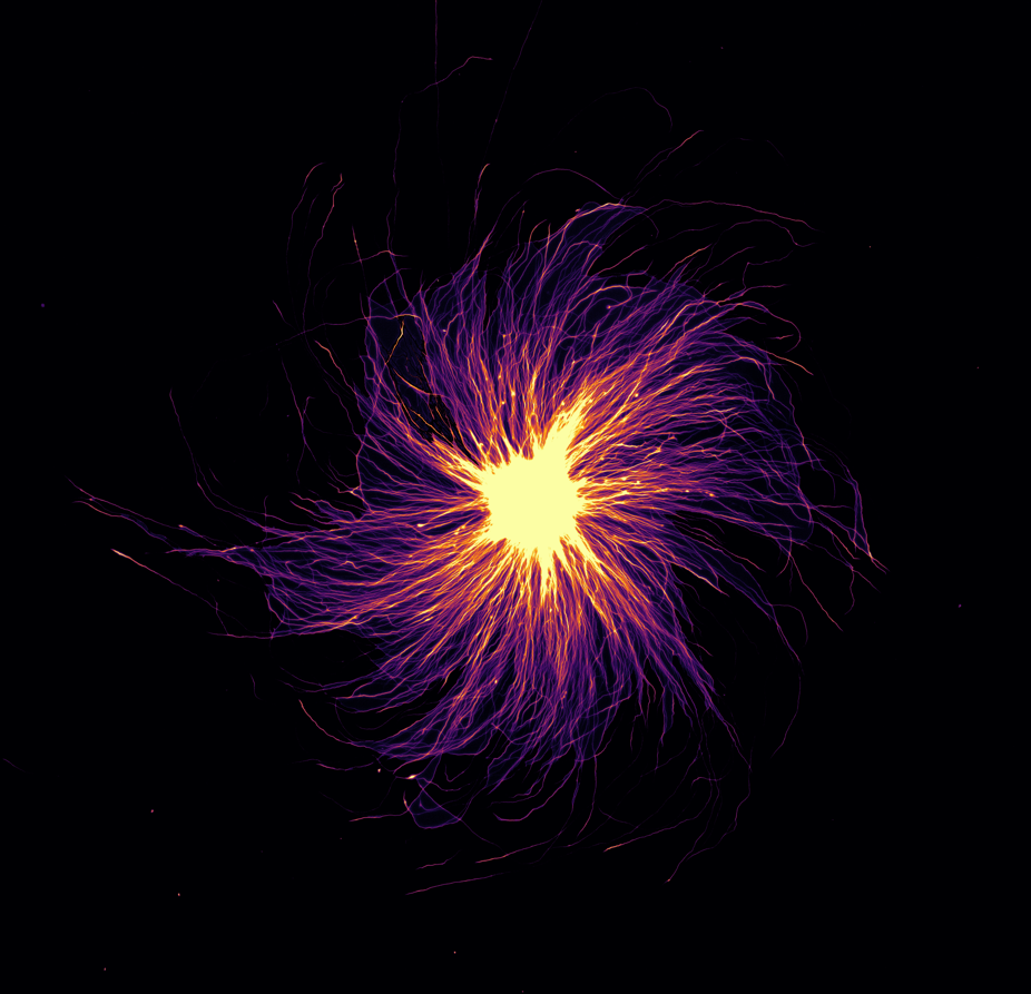
Centre for Craniofacial & Regenerative Biology, Faculty of Dentistry, Oral & Craniofacial SciencesAxons of human iPSC derived motorneurons growing out of a neurosphere
Stem cell models allow us to recapitulate neurodevelopment in vitro and to investigate in depth the biological processes underlying neuronal function and neurodegeneration. However, conventional cell culture does not always resemble the highly specialised structure of neurons such as motorneurons. These cells establish long ranged connections with axons spanning over 1m in humans. Modelling axonal length would allow us to understand the specific constrains associated with length in health and disease. We develop a device to systematically investigate axonal length in vitro.
Therefore, we are investigating the growth behaviour of axons on different substrates. This image was taken in an experimental series to investigate the interaction between motorneuron-spheres and their substrate, in this case a silicon-based polymer. The neurons in this image were cultured for 7 days on the substrate and then antibody stained for beta-3-tubulin, a prominent axonal marker. This image was taken with a fluorescent microscope at 20x magnification and post processed in ImageJ using false colouring.
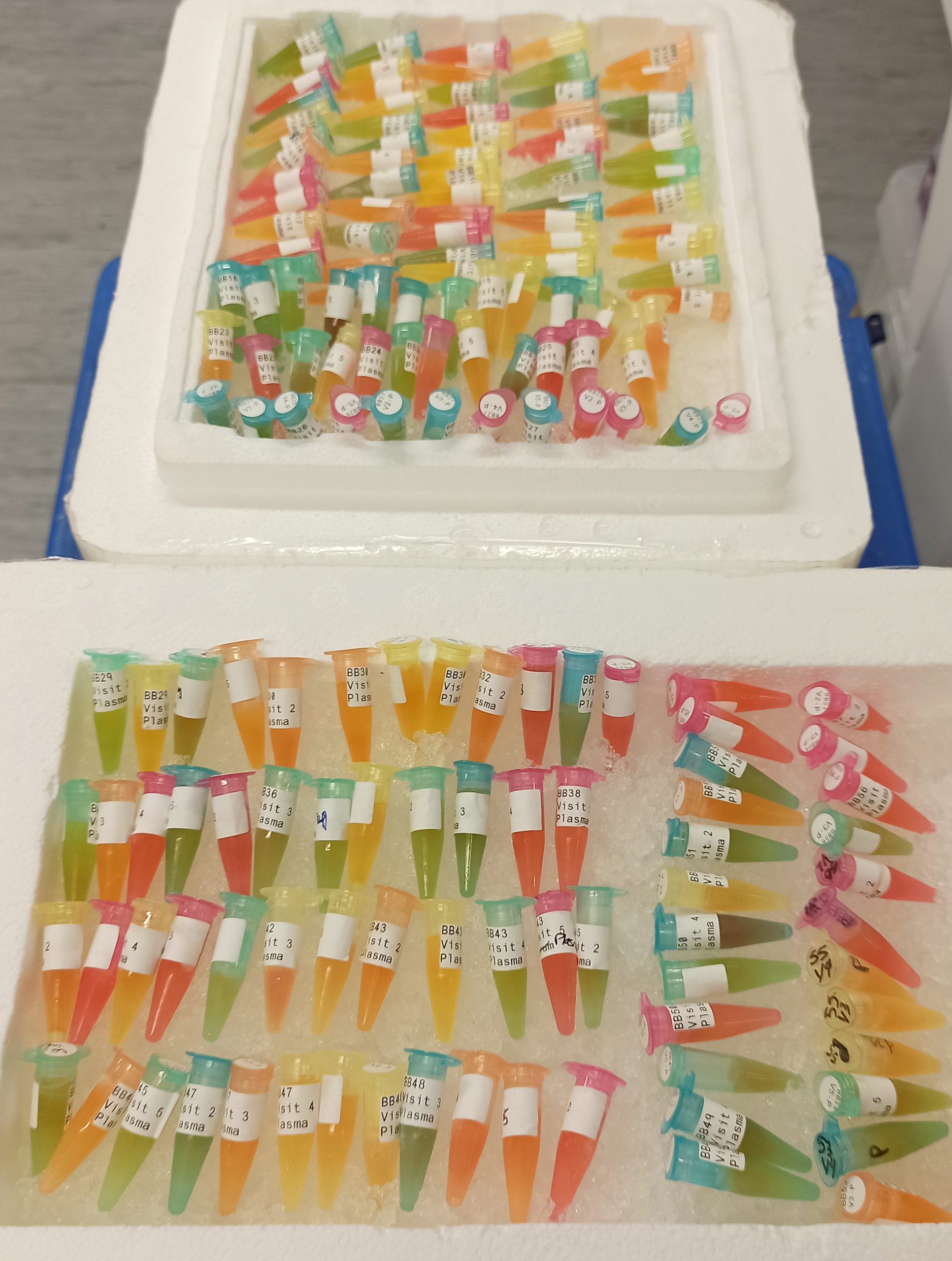
Department of Nutritional Sciences, Faculty of Life Sciences & MedicinePut some rainbows in your research!
Plasma samples being thawed on ice, in anticipation for sample preparation and analysis. Plasma samples have been previously collected as part of a human clinical trial investigating the impact of blueberry consumption on cardiovascular parameters. The purpose of this analysis is to identify and quantify specific molecules called (poly)phenols – present in blueberries – in the plasma. The final aim would be to understand the metabolism of blueberries following their ingestion. Photo (unmodified, only cropped) taken on the 26th of February 2021, in FWB (Waterloo campus).
Nicholas Merrild (shortlisted)
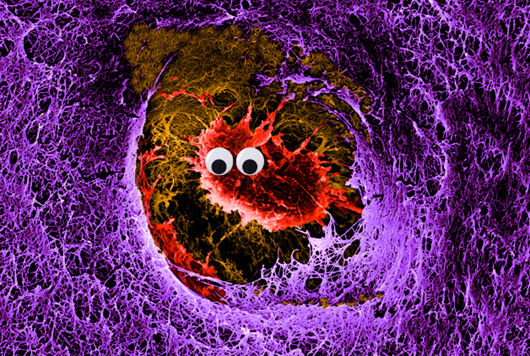
Centre for Craniofacial & Regenerative Biology, Faculty of Dentistry, Oral & Craniofacial SciencesWhen the Void Stares Back
Comprised of a web of highly dense, organised and beautiful rope-like structural proteins, collagen carries and distributes the tension of the loadbearing connective tissue of joints-articular cartilage (AC). If adult AC is fractured, or otherwise lacerated, regeneration is unlikely in adults and the tissue’s further decline inevitably leads to a condition known as osteoarthritis. What little repair can be done, is managed by the sole cell type found in AC –the chondrocyte. However, chondrocytes only comprise 5-10% of the tissue volume and live isolated lives in their lacunae –literally, a well–trapped in a dense matrix of the rope-like collagen fibres.
Using a juvenile pig AC wound model in vitro, we can successfully observe repair after injury. To understand the injury response, we can image the dense collagen network surrounding the cell. Having ruled out the generation of new collagen, collagen cross-linkers, or any macro molecules generally associated with aiding cross-linking, we stumbled upon the absurd observation that a protein generally only associated with advancing osteoarthritis, may play an active role in repair. Upon confirming its importance, we aim to leverage this protein for tissue engineering strategies. So, after years of staring into the deep dark void of the well, it now stares back at us…and it is absurd.
Using scanning electron microscopy, this image (width just under 25μm; ~1/40th of a millimetre) shows a single chondrocyte embedded in a dense nest of collagen fibres averaging ~100nm (~1/10,000thof a millimetre!) in diameter. The original black/white image was false coloured in photoshop, and brightness and contrast were adjusted, and finally googly eyes were added to produce the final image submitted.
Disclaimer: googly eyes are not naturally found on chondrocytes, added for artistic purposes only.
HSDTC Science Communication Competition 2021
PhD students undertook the challenge of submitting a short ‘newspaper style’ article about a research project they were currently involved in (or an aspect of it). The article is aimed at a non-expert audience and had to be engaging and understandable for an interested member of the public.
The judges were looking for articles which: are compelling to read and easily understandable; clearly explain the research being done; answer the question “why does this research matter?”; and are worthy of publication in a national newspaper.
Five fascinating submissions were shortlisted, including the winning submission by Elisa Brann, and the runner-up, Simon Lam:
Forensic & Neurodevelopmental Sciences, Institute of Psychiatry, Psychology & NeuroscienceA little magic in science: exploring psychosis using hypnosis
Psychosis is a devastating condition that involves perceiving things that are not really there (hallucinations) or believing things that are not really true (delusions). Approximately 5% of people who experience psychosis commit suicide, making it one of the deadliest psychiatric conditions you can have. Whilst it is usually treated using anti-psychotic medication, sadly, one in four people do not see improvements.
One way of developing new therapies for psychosis is by understanding what is happening in the brain as it happens. But conducting brain imaging on these individuals is no simple task. Someone in the midst of a psychotic episode is usually far too unwell to take part in research. How then can we ever understand what’s going on?
Researchers at King’s College London have come up with an innovative way of tackling this issue – they conduct their brain imaging on healthy people who have been hypnotised to have psychotic experiences.
Hypnosis describes a scenario in which an individual enters a subjective state where alterations of perception can be caused by verbal instructions, known as suggestions. Many people are sceptical about hypnosis, often associating it with magic. But it is a very real phenomenon that can result in very powerful experiences. It has a long history of being used as an alternative to traditional anaesthetics, even during brain surgery!
By carefully crafting suggestions for a select group of people who are particularly hypnotizable, the researchers at King’s College London have been able to create many different kinds of psychotic symptoms. These include auditory verbal hallucinations (‘hearing voices’), delusions of control (when you believe someone else is controlling your body) and thought insertion (when you believe someone else is putting thoughts in your mind).
Now that they have been able to reliably produce these experiences, the research team are beginning to document what is happening in the brain as they happen. They hope that in doing so they will open a window into how psychosis works and ultimately unlock new ways of treating it. Sometimes you need to think outside the box in science, sometimes you need a little magic.
Centre for Host-Microbiome Interactions, Faculty of Dentistry, Oral & Craniofacial SciencesFishing for an Alzheimer’s cure – how researchers use zebrafish to study the brain
It turns out that fish really is good for your brain – but not how you might think.The common adage that fish is brain food is undisputed. It is well established that eating fish and taking fish oil capsules are good for the brain. But researchers have found another way that our scaly friends help our brains, and it doesn’t involve eating them.
Scientists studying Alzheimer’s and Parkinson’s at King’s College London have found that old age affects zebrafish brains in similar ways as it does humans. They found that as the zebrafish age, the amount of retinoid in the brain decreases. This is the same as what they observed in Alzheimer’s and Parkinson’s patients.
“Retinoids are molecules associated with anti-ageing,” explains Simon Lam, a second-year PhD student at the Centre for Host-Microbiome Interactions. “We get them through our diet by eating foods like carrots and broccoli. But they’re also found in anti-ageing formulas and acne creams.”
It might not seem too surprising that known anti-ageing molecules should crop up when studying ageing, says the group. But it does help unlock the link between brain ageing and Alzheimer’s and Parkinson’s.
“And that’s what makes this finding so exciting. Old age is a risk factor for Alzheimer’s and Parkinson’s, but it’s unclear how that works. What we’re trying to do here is tease out the changes in our brain in old age, and that’s why we’re using the zebrafish. We have fast and slow ageing fish, which should be able to tell us more about those changes.”
But when asked if eating more carrots is the cure to Alzheimer’s, the team says it isn’t so simple.
“What makes Alzheimer’s and Parkinson’s so difficult to treat is that patients have highly variable responses to treatment. A treatment that works on one patient may or may not work on another. These are open questions we’re trying to answer: can we form groups of patients for which a specific treatment will work? And can we sort patients into the right group?”
Centre for Craniofacial & Regenerative Biology, Faculty of Dentistry, Oral & Craniofacial SciencesJust go with the flow! Microfluidics in cell therapy production.
Cell therapy is a revolutionary concept in medical science, whereby living cells are administered to an individual, often by injection, in order to treat a specific disease. An example of this is CAR T-cell therapy, where cells of the immune system are extracted from the blood of a cancer patient and genetically edited to recognise cancer cells. They are then re-infused back into the patient where they will target and attack malignant tumours.
CAR T-cell therapy was approved in the UK in 2017 and has since been touted as one of the most important advances in cancer research in the 21st century. Despite its ability to induce complete remission in up to 90% of patients, it is currently not used as a first-line treatment as a result of the high cost for the NHS. The complex multi-step manufacturing process, requiring specialist equipment and expensive reagents, is reflected in the high price-point of £280,000 per individual.
In order to drive down CAR T-cell therapy manufacturing costs, researchers at King’s College London are attempting to use microfluidic technology in the CAR T-cell manufacturing process. Defined as the manipulation of fluids on a micro-scale, this technology has the potential to signficantly reduce costs by processing cells using a fraction of the reagents that are required with conventional methods. In addition, microfluidic approaches could also provide a higher degree of process stability and control. Typically, around 10% of patients who are eligible for the treatment do not receive it, as a result of errors in the manufacturing process. By allowing the automation of certain processes, this approach could reduce variability and human-related error, resulting in a better-quality product.
With the ambition of developing a single hand-held device to perform all of the operations of an entire laboratory required for the manufacturing process, this technology could overcome financial barriers and ultimately make CAR T-cell therapy accessible to more individuals.
Department of Nutritional Sciences, Faculty of Life Sciences & MedicineAronia: a “berry” good ally for your health!
Have you ever heard of aronia berry, a fruit coming from North America and looking like a small dark blueberry? Probably not, yet this berry is becoming of interest in the nutritional research field. Indeed, due to its high concentration in (poly)phenols – a family of molecules present in all plant-based foods and demonstrating benefits on various chronic diseases including cancer – numerous human trials have decided to investigate the potential of aronia on cardiovascular health. Indeed, several experiments have revealed the ability of aronia (poly)phenols to significantly improve blood pressure and cholesterol levels following daily consumption of aronia extract.
Recently, a study led by researchers from King’s College London examined the impact of the berry on arterial stiffness, a parameter increasing naturally with age and related to the rigidity of our blood vessels. Their study consisted in a human trial involving more than 100 healthy middle-aged men and women who took 1 capsule per day for 12 weeks, containing either aronia (poly)phenols or a placebo. Investigators performed some measurements including arterial stiffness at baseline and after the intervention, and they compared the results to evaluate the effects of the berry extract. After 12 weeks of intake, they observed that subjects consuming aronia (poly)phenols had significantly decreased arterial stiffness, implying an improvement of their cardiovascular health.
As cardiovascular diseases account for more than 25% of the overall deaths in the UK each year, the beneficial effects of aronia berry on vascular health could help relieve the socio-economic burden associated to the incidence of these pathologies. Besides, although aronia berries are not widely available in the UK yet, the same beneficial (poly)phenols can be found in various foods, from the blueberries you have for breakfast to a satisfying glass of red wine. A great excuse to increase your intake in (poly)phenols!
Centre for Host-Microbiome Interactions, Faculty of Dentistry, Oral & Craniofacial SciencesFighting a global health threat: fungal infections
Fungal infections affect 1 billion people worldwide and kill 1.5 million individuals every year. Yet, they are still extremely underestimated and understudied. Currently, there are no vaccines against fungal infections.
The most common human fungal infections are nail & skin infections, and thrush. They are treatable with antifungals, but can be distressing and there is no preventative treatment. Vaginal thrush is a major problem: 3 out of 4 women will suffer from it at least once in their lifetime. Many will also experience painful recurring episodes.
The main cause of thrush? The fungus Candida albicans.
Candida albicans is also responsible for severe bloodstream infections in people with a weakened immune system and hospitalised patients. These are extremely challenging to treat and reach mortality rates of 50%.
So, what kind of research is being done?
In 2016, researchers from the Naglik laboratory at King’s College London discovered a toxin produced by Candida albicans. The toxin physically and chemically damages human cells during an infection and causes inflammatory symptoms. They named it ‘candidalysin’.
Since then, research has been focused on understanding the exact mechanisms by which candidalysin damages human cells. By investigating how candidalysin works, we may be able to find ways to block its activity and thus decrease Candida albicans’ ability to cause diseases.
What are the next steps?
The discovery of candidalysin creates a new field of science, by being the first toxin of its kind to ever be discovered in a disease-causing fungus. Further research will allow us to better understand fungal diseases, and to develop novel diagnoses and therapies. Most importantly, this could also lead to the development of one of the first fungal vaccines ever.
Understanding candidalysin gives us hope to significantly reduce the suffering and death of millions of people worldwide.